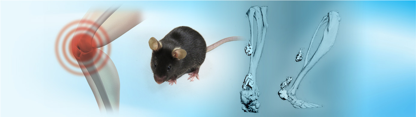
Heterotopic ossification (HO) is abnormal bone formation in soft tissues. It can be genetic or non-genetic. Non-genetic HO is caused by surgery, trauma, or thermal injury and leads to pain, swelling, and joint immobility. It can occur either locally or at distant, uninjured sites, making targeted interventions challenging. This case study demonstrates the expertise of Aragen’s scientists that helped its client in surmounting the challenge of unpredictable HO localization by reproducing the thermal lesion/tenotomy-induced mouse model. The model recreates local trauma (tenotomy) and global inflammation (distant thermal lesion) simultaneously, producing focussed HO and, faacilitating the development of targeted interventions for HO.
Here, at Aragen, our scientists have characterized thermal lesion/tenotomy induced mouse model of non-genetic heterotopic ossification. Local HO was developed by an Achilles’ tenotomy on the left hindlimb, followed by thermal lesion on the dorsal skin. Post-surgery, the animals were weighed every alternate day and observed for clinical symptoms during the study duration. After 62 days of thermal lesion/tenotomy, tissues (hindlimbs) were collected and submitted for Micro CT acquisition and analysis. All experimental protocols adhered to strict ethical standards and received regulatory approvals, ensuring compliance with guidelines governing animal model studies.
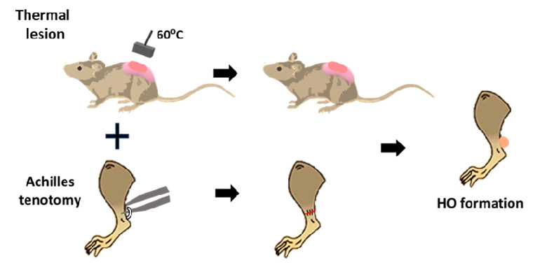
Figure 1: Study design.
Aragen’s scientists successfully reproduced the thermal lesion/tenotomy-induced mouse model that reliably generated heterotopic ossification at the site of direct trauma in all the experimental animals. In addition to its reliability, this model offers the advantage of HO formation predictability via its ability to induce ectopic bone formation in response to trauma/thermal lesion.
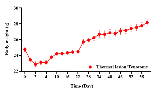
Figure 2: A decrease in the body weight of experimental mice was observed for the initial seven days post thermal lesion/tenotomy injury, followed by an increase in their body weight thereafter.
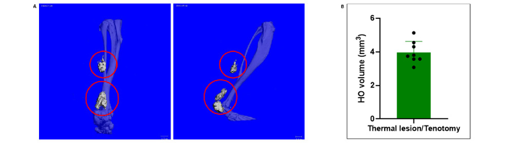
Figure 3: Micro CT images of the tenotomy site (sagittal view) depicting heterotopic ossification sites (red circles) following tenotomy/dorsal skin thermal lesion (A); Heterotopic ossification volume measurement (B).
Our experienced staff with more than 15 years of expertise provide:
We extend our sincere appreciation to Charles River Laboratories for their invaluable contribution to Micro CT acquisition and analysis. Additionally, we would like to acknowledge Benjamin Levi and his esteemed team at the Department of Surgery, University of Michigan Medical School, for establishing the model, which we simulated in the current study [Peterson, J.R., Agarwal, S., Brownley, R.C., Loder, S.J., Ranganathan, K., Cederna, P.S., Mishina, Y., Wang, S.C., Levi, B. (2015). Direct mouse trauma/burn model of heterotopic ossification. J. Vis. Exp. (102), e52880, doi:10.3791/52880].